|
Heart Rate Variability and Pulse wave form – Diagnose body (constitution) phenotype:
There are doctors in India, China and Tibet that have developed the art of "listening to the music ofthe heart," over thousands of years and they are very good at it. There is ongoing research in Russiawhere researchers are decoding the pure sound of the heart working with an unpolluted, cleansignal.
The ancient doctors used the pulses as the reflections of the original impulses of the heart. Pulse diagnosis is one of the most important diagnostic methods in traditional East Asian medicineBy diagnosing the pulse, trained practitioners can reveal information on the balanceof Qi and blood and on the homeostasis of the body and various organ systems. Historically, thepulse was assessed at various regions of the body, including the carotid artery, temporal artery,dorsalis pedis, femoral and popliteal arteries, and radial arteries. In contemporary traditional East Asian medicine, thepulse is commonly diagnosed using the radial arteries of both wrists.
Thesepoints are called Chinese Cun, Guan, Chi.(cun guan chi (inch, gate, foot)) Ayurved medical system Tridosha i.e.Vata(catabolic), Pitta(metabolic) and Kapha(anabolic) They are resonant with thefrequencies given by the heart. Depending on what point is palpated by finger pressure and at whatdepth, the state of the organ or system corresponding to that point can be judged.
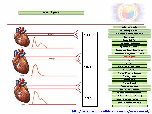
These regions of the wristcomprise three distinct palpation locations that span 5 cm along the radial artery: Cun (Chon),Guan (Gwan), and Chi (Cheok).It is said the Cuan position corresponds to the region of the body from the bottom of the chest tothe top of the head;the Guanposition corresponds to the area of the body located between the diaphragm and the navel; and theChi position corresponds to the region of body that is below the navel, Pulse signals measured from Chon (Cun)- Vata, Gwan (Guan)-Pitta, and Cheok (Chi)-Kaphawhich are three adjacentregions along the radial artery used for pulse diagnosis in traditional Eastern medicine (TEM), canreflect a patient's physical condition and pathological problems. Therefore, pulse diagnosis hasbeen considered an important diagnostic method in traditional systems of medicine for thousands of years.The balance of the pulses felt between Cun, Guan, and Chi reflects the balance between thecorresponding organ functions in the upper, middle, and lower body parts. Similarly, the pulsesbetween the left and right wrists describe the balance between the left and right parts of the body.
Thus, the pulse images felt at the cun position indicate the condition of the heart and lungs, those at
the guan position that of the liver and spleen, and those at the chi position that of the two kidneys
(or the kidneys and the “gate of life” or yet another variant)the three sectors cun, guan, chi correlate again with the upper, middle and lower burner
Upper burner
In this section the heart, corresponding with the small intestine on the left hand side, and the lungs,
corresponding with the large intestine on the right hand side, are said to unite in the upper burner;
Middle burner
the liver, corresponding with the gall bladder on the left hand side and the spleen, corresponding
with the stomach on the right hand side, are said to unite in the middle burner;
Lower burner
the kidneys onthe left and right hand side, corresponding with the bladder, and the “portal of the womb” ( baomen C ) on the left or the “door to the child” ( zi hu E ) on the right hand side are said to unitein the lower burner (cols 135 and 137)
It is said the Cuan position corresponds to the region of the body from the bottom of the chest to
the top of the head;the Guanposition corresponds to the area of the body located between the diaphragm and the navel; and theChi psition corresponds to the region of body that is below the navel


For instance, the pulse at Cun describes the organ function within the upper region of the trunk and
thoracic cavity, such as that of the lungs and heart; the pulse at Guan describes the condition of the
upper abdominal cavity, which includes the liver, spleen, and pancreas; and the pulse at Chi reflects
the condition of the lower abdominal cavity, which includes the urinary and reproductive organs.
The balance of the pulses felt between Cun, Guan, and Chi reflects the balance between the
corresponding organ functions in the upper, middle, and lower body parts. Similarly, the pulses
between the left and right wrists describe the balance between the left and right parts of the body.
Pulse diagnosis is a palpation method that is one of the four diagnostic examinations of Korean
medicine. It is a diagnostic method in which a doctor uses finger sensations to observe the pressure
pulse waveform (PPW). Korean medical doctors consider this PPW information to understand a
patient's health condition or validate medical treatment. In the early days, doctors sensed PPWs at
various sites over the whole body, but this has changed to the wrist pulse-taking method. Pulse
diagnosis has very high diagnostic importance and significance in Korean medicine, but it depends
on a doctor's subjective sensations and oral tradition. To overcome these issues, many studies have
been conducted on the objectification and quantification of pulse diagnosis, and electromechanical
pulse diagnostic devices have been invented.
Pulse Pressure Wave analysis with Fourier transformandwavelet transform,and respiration rate extraction using fast Fourier transform have beenconductedPulse is one of the most critical signals of human life. It comes directly from heart to the bloodvessel system. As pulse transmitted, reflections will occur at different level of blood vessels. Otherconditions such as resistance of blood flow, elastic of vessel wall, and blood viscosity have clearinfluence on pulse. Pathological changes affect pulse in different ways:the strength, reflection, and frequency. So pulse provides abundant and reliable information aboutcardiovascular system. Advanced Biomedical Engineering correlated with Pulse Wave Velocity -PWV (r=0.65, P<0.0001).It is an effective non-invasive method for assessing artery stiffness.Pulse Wave Velocity is the golden standard for arterial stiffness diagnosis. Researches show thatStiffness Index has equivalent output as PWV.
Pulse diagnosis a diagnostic method in which a doctor uses finger sensations to observe the pressurepulse waveform (PPW). Traditional Eastern medical practitioners consider this pulse waveform information to understand apatient's health condition or validate medical treatment. Pulsediagnosis has very high diagnostic importance and significance in eastern medicine, but it dependson a doctor's subjective sensations and oral tradition. To overcome these issues, many studies havebeen conducted on the objectification and quantification of pulse diagnosis, and electromechanicalpulse diagnostic devices have been invented.
The arterial pulse waveform has been widely used to diagnose and treat various diseases The
Eastern medical system of pulse feeling has a history of more than 2,000 years. According to Traditional ChinesePulse Diagnosis (TCPD) that provides a convenient, painless, and noninvasive diagnosis method, wecan deduce the position and degree of pathological changes from pulse waveform. Accordingto the theory of modern medicine, the pressure wave in radial artery at wrist provides anestimation of arterial pressure wave in the proximal aorta. It can reveal central systolic, diastolicand mean arterial pressures, as well as supply an assessment of arterial wave reflection, which isclosely related to cardiovascular condition and the degree of artery stiffness. Pulse interval variability(PIV), which refers to the beat-to-beat alterations in pulse rate, is essentially similar to Heart RateVariability. The variability is mainly due to the control thatthe autonomic nervous system exerts on the heart.The arterial pulse waveform is a contour wave generated by the heart when it contracts, and ittravels along the arterial walls of the arterial tree. Generally, there are 2 main components of this
wave: forward moving wave and a reflected wave. The forward wave is generated when the heart(ventricles) contracts during systole. This wave travels down the large aorta from the heart and getsreflected at the bifurcation or the "cross-road" of the aorta into 2 iliac vessels.
Pulse is one of the most critical signals of human life. It comes directly from heart to the bloodvessel system. As pulse transmitted, reflections will occur at different level of blood vessels. Otherconditions such as resistance of blood flow, elastic of vessel wall, and blood viscosity have clearinfluence on pulse. Pathological changes affect pulse in different ways:the strength, reflection, and frequency.Pulse Pressure Wave analysis with Fourier transformandwavelet transform,and respiration rate extraction using fast Fourier transform have beenconducted by several studies.
Pulse Wave Velocity -PWV is an effective non-invasive method for assessing artery stiffness.Pulse Wave Velocity is the golden standard for arterial stiffness diagnosis. Researches show thatStiffness Index has equivalent output as PWV.
It uses the reflection of the pulse as a source to get the time difference without additional sensors which make it more applicable to theHome Monitoring System. The systolic component shows the time that pulse reach thefinger; diastolic component represents the time that pulse reflection reach the finger. The first part of the waveform (systolic component) is result of pressure transmissions along a directpath from the aortic root to the finger. The second part (diastolic component) is caused by thepressure transmitted from the ventricle along the aorta to thelower body. The time interval between the diastolic component and the systolic component dependsupon the Pulse Wave Velocity of the pressure waves within the aorta and large arteries which is related to arterystiffness. PWV is the velocity of the pulse pressure. Total arterial compliance and increased central Pulse Wave Velocity (PWV) are associated with arterial wall stiffening. PWV is the velocity of the pulse pressure. Stiffness Index (SI), which is derived from the pulse wave analysis for artery stiffness assessment and was correlated with PWV. It is an effective non-invasive method for assessing artery stiffness.The arterial Stiffness index is an estimate of the pulse wave velocity (PWV)about artery stiffness and is obtained from subject height(h) divided by the time between the systolic and diastolic peaks of the pulse wave contour. Theheight of the diastolic component of the pulse wave relates to the amount of pressure wavereflection.Stiffness index is related to the time delay between the systolic and diastolic componentsof the waveform and the subject’s height. SI is highly related to the pulse rate because it is calculatedby the time interval between systole and diastole. Younger people with high pulse rate can get arelative high score than older people with slow pulse rate. Adjustment based on pulse rate can beapplied on SI calculation.Fourier Transform and Wavelet Transform have been used toperform the basic analysis onthe pulse waveform. Fourier transform is a basic and important for linear analysis whichusually transfers the signal from time domain (signal based on time) to frequency domain (thetransform depends on frequency). The Fourier theory states that any continuous signals or timeserial data can be expressed as overlay of sine waves with different frequencies. This process canhelp signal analysis because the sine wave is well understood and treated as simple function in bothmathematics and physics. Fourier transform calculates the frequency, amplitude, and phase based onthis theory.
The autonomic nervous system (ANS) regulates individual organ function and homeostasis, such asheartbeat, digestion, breathing and blood flow, and for the most part is not subject to voluntarycontrol. These involuntary actions are controlled by the opposite actions of the two divisions of theautonomic nervous system—the sympathetic and the parasympathetic divisions. Most organsreceive impulses from both divisions and under normal circumstances and they work together forproper organ function and adaptation to the demands of life. Problems will occur when theautonomic nervous system is out of balance, for example, coronary heart disease, hypertension,digestive disturbances and even sudden death.The autonomic nervous system ANS (visceral nervous system or involuntary nervous system ) isthe part of the peripheral nervous system that acts as a control system that functions largely belowthe level of consciousness to control visceral functions, including heart rate, digestion, respiratory rate, salivation, perspiration, pupillary dilation,micturition (urination), sexual arousal, breathing and swallowing. Most autonomous functions areinvoluntary but they can often work in conjunction with the somatic nervous system which providesvoluntary control.The ANS is divided into three main sub-systems: the parasympathetic nervous system (PSNS),sympathetic nervous system (SNS),and the enteric nervous system (ENS). Depending on the circumstances, these sub-systemsmay operate independently of each other or interact co-operatively.
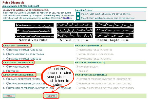
Pulse diagnosis (Nadi Pariksha- Indian Ayurved medical system) is done to analyze and estimate the quantity of Tridosha (phenotype/constitution) in the body. Tridosha i.e.Vata, Pitta and Kapha areconsidered as the fundamental elements of health. A balance between these three is considered asPrakruti or healthy statusand any imbalance in these three is considered as Vikruti or ill health. As per ancient Ayurvedictext, pulse (Nadi) can beexamined at various places but commonly it is examined at the wrist of the person.
It is observed that apart from the underlying disease, the Dosha dominance of an individual changeswith age. Forinstance Kapha is more dominant in the childhood, Pitta is more dominant in the middle age andVata is dominant in oldage. (In present times Pitta dominance is seen in twenties and thirties and Vata dominance is seenbeyond forties.) .
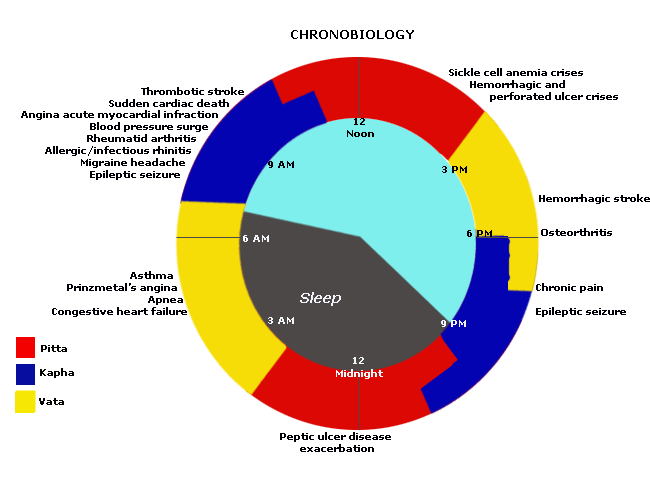
Also rhythmic variations are found in these Dosha in an individual during a day.The observation that HighFrequency peak in HRV spectrum goes on reducing as the age advancesand may touch the base line in cases of severe illness suggests that strength of the HighFrequency peak mayhave inverse relation with the level of Vata element in the body. Similarly mid/low Frequency may have direct relation with Pitta levels in the body.The shape or pattern (morphology) of the peripheral pulse changes significantly in an individual as afunction of time.
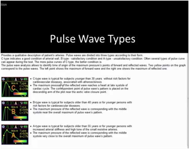
Pulse Waveform of a younger person(Cheok (Chi)-Kapha)
For a normal young person, where the arteries are generally compliant, the slow travelling reflectedwave from the peripheral occurs during diastole, thus enhancing perfusion of the coronary arteries.
Pulse Waveform of a middle aged person(Gwan (Guan)-Pitta)
With age, the arteries stiffen. Pulse wave velocity increases and the reflected wave now travels fasterand gives rise to a "shouldering" at the primary wave, and occurs during systole. This effect prolongsthe systolic cycle increasing the workload and oxygen requirement of the heart muscles. The poornotch also compromises the flow of the coronaries.
Pulse Waveform of an elderly person(Chon (Cun)- Vata)
In the elderly, further arterial stiffening cause the reflected wave to return much faster and gives riseto a "shouldering" at the primary wave; and coincide very close to the systolic peak, resulting in anaugmented wave. The heart now needs to contract even harder (to overcome the oncomingreflected wave) and for a longer period. At the same time, coronary artery perfusion is furthercompromised.
Patterns of heart rate variation relate closely to the modulation of the autonomic nervous system.Heart rate variability (HRV) has been developed as a function indicator of the autonomic nervoussystem. HRV refers to the beat-to-beat alterations in the heart rate. It is a measure of thebeat-to-beat time interval variations as the heart speeds up or slows down with each breath under aprecordial state. Among the different techniques in assessing the autonomic nervous system, HRV isan important breakthrough because this technique is non-invasive. Moreover, the hardware for thetechnique is inexpensive, and thus can be broadly applied In addition, animal and clinical studies confirmHRV accurately reflects the sympathetic and parasympathetic activities and their balance.Electrical engineering have enabled the assessment of heart rate variability by frequency domainanalysis, which is based on mathematical manipulations performed on the ECG-derived data.
A pulse wave form is first taken from a subject and then amplified,filtered and digitized. A computer program for HRV analysis is then used to process the ECGsignals. The computer first detected all peaks of the digitized ECG signals. The interval between twopeaks is then estimated. The frequency-domain measurements are further quantified by using anonparametric method of fast Fourier transform (FFT).Based on frequency analysis, HRV can be characterized into two main components: the highfrequency (HF) component and the low frequency (LF) component. The high frequency componentis equivalent to respiratory sinus arrhythmia and is considered to represent the influence of the vagalcontrol of the heart rate- Parasympathetic-Kapha. The exact origin of the low frequency component is not known. It isprobably related to vessel activity or baroreflex-sympathetic.activity-Pitta. Some investigators further divide the low frequencycomponent into a low frequency component and a very low frequency component.- enteric nervous system (ENS)- Vata
Continuous Impedance plethysmography (IPG)signal is recorded from a body segment for a period of two-five minutes. Blood flow index values are then obtained from this signal as a function of time and interpolated to getequi-spaced values. Fourier Transform of this time series then gives the rhythm of fluctuations.The peak at 0.012 Hz. represents activity of thermo-regulation/ Baro-receptor reflex/sympathetic nervous system respiration respectively.High frequency component or the total power (TP) can consider representing the parasympatheticcontrol of the heart rate and the ratio LF/HF is considered to mirror the sympathovagal balance orto reflect the sympathetic modulations, very low frequency component- enteric nervous system (ENS).Smartphone microphoneis placed near the heart of the subject to collect two to five minutes ofthe sound signals of the heart and index finger is placed on the camera(LED light of the smartphone) The sound and light signals of the heart are amplified, filtered and transmittedto an analogy-to-digital (A/D) converter.The digitized sound/light signal is then analyzed to determine the beat-to-beat interval using a computerized analysis.Parameters such as amplitude and duration of all peaks are determined so that their means andstandard deviations are calculated as standard templates. Each subsequent heart rate is thencompared with the standard templates. The power spectral density is further estimated on the basisof fast Fourier transform and is subsequently quantified by means of integration into standardfrequency-domain measurements including very low frequency(VLF), low frequency (LF), high frequency (HF), total powerand LF/HF. Then these parameters are logarithmically transformed to assess body phenotype(constitution)


|